Distinguishing lesions from posterior acoustic shadowing in breast ultrasound via non-linear dimensionality reduction. Posterior acoustic shadowing Non-circumscribed margins Non-palpable Controversial and age dependent Not new Case 3.
Basic Principles Radiology Key
The cases illustrate a variety of pathologic breast conditions that were collected at a referral breast center at a tertiary medical center.

. Breast Ultrasound Past. Multiple projections from the nodule within or around ducts extending away from the nipple usually seen in larger tumors. In this paper half-contour features are proposed to classify benign and malignant breast tumors with PAS considering the fact that the upper half of the tumor contour is less affected by PAS.
It is wider than tall with macrolobulations no calcifications and posterior acoustic shadowing. The phenomenon of acoustic shadowing sometimes somewhat tautologically called posterior acoustic shadowing on an ultrasound image is characterized by a signal void behind structures that strongly absorb or reflect ultrasonic waves. Posterior acoustic features are described as no posterior acoustic features enhancement white shadowing dark or a combined pattern.
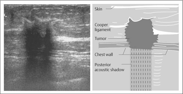
Acoustic shadowing originates from Coopers ligament between normal fat lobules. The results of pathology pending. The boundary between the mass and the surrounding tissue is described as having an abrupt interface or as containing an echogenic halo a white blurry band surrounding the mass.
Breast sonogram reveals focus of intense acoustic attenuation without mass lesion. Shadowing may result because of reflection of most of the energy by a large impedance discontinuity. Download scientific diagram Transverse ultrasound of the left breast demonstrates an irregular antiparallel mass with posterior acoustic shadowing.
It is a form of imaging artifact. 70 F remote history of breast cancer and prior lumpectomy. Which usually do not shadow.
Up to 20 cash back I had a breast ultrasound with core biopsy. Everything I read points towards breast cancer. Screen detected new mammographic mass Ultrasound is planned as the next step for this finding.
Breast ultrasound US in conjunction with digital mammography has come to be regarded as the gold standard for breast cancer diagnosis. Sonographic posterior acoustic shadowing. With remarkably intense and sharp posterior acoustic shadowing Fig.
Posterior acoustic shadowing PAS can bias breast tumor segmentation and classification in ultrasound images. Variable sized fat deposits surrounded by foamy. Developing asymmetry upper inner breast mid depth.
Theultrasound showed a 40 x 24 x 40cm irregular mass with angular margins posterior shadowing and architecturl distort. Am having stereotactic biopsy this week. It is a form of imaging artifact.
AbstractBreast ultrasound US in conjunction with digital mammography has come to be regarded as the gold standard for breast cancer diagnosis. I had core needle biopsy which came back as benign breast tissue. A variety of pathologic conditions are discussed with pathologic-imaging correlation.
Hypoechoic irregular mass with posterior shadowing indistinct margins and vascular flow measuring 11 x 4 x 9 mm. It is the posterior acoustic shadowing that is freaking me out. While breast US has certain advantages over digital mammography it suffers from image artifacts such as.
An irregular hypoechoic mass with intense posterior acoustic shadowing can be typically seen on US and can mimic breast malignancy Fig. This loss is displayed in the image as shadowing and is an important sonographic sign for the detection and diagnosis of breast disease. You will be shown possible ultrasound correlates on the next slide and be asked to pick the best correlates and next step.
The presence of a foreign body at sonography is easily con-firmed by correlation with mammograms. Type I is a hyperechoic semilunar structure with posterior acoustic shadowing type II is a curvilinear echogenic structure with acoustic shadowing and type III are irregular clumps. While breast US has certain advantages over digital mammography it suffers from image artifacts such as posterior acoustic shadowing PAS presence of which often obfuscates lesion margins.
DMP usually shows nonspecific parenchymal enhancement rather than an irregular enhancing mass on MRI. Acoustic shadowing from Coopers suspensory ligament mimics breast neoplasm at sonography of normal breast in 43-year-old woman. On mammography the lesion usually shows localized increased density in the glandular tissue.
C Ultrasound of the left breast shows a 17 mm lesion which appears completely hyperechoic but with suspicious features including an ill-defined margin and posterior acoustic shadowing. The phenomenon ofacoustic shadowing sometimes somewhat tautologically called posterior acoustic shadowing on an ultrasound image is characterized by a signal void behind structures that strongly absorb or reflect ultrasonic waves. A small amount of extracapsular silicone due to implant rupture may initially be mis-taken for a cyst or hypoechoic solid mass with posterior acoustic enhancement Fig.
Although posterior acoustic shadowing is a sonographic feature that is most commonly associated with mammary. Am petrified Has anyone had posterior acoustic shadowing on a. As ultrasonic beams propagate through tissues there is a loss of energy by absorption reflection and scattering.

Posterior Acoustic Shadowing In Benign Breast Lesions Weinstein 2004 Journal Of Ultrasound In Medicine Wiley Online Library

Posterior Acoustic Shadowing In Benign Breast Lesions Weinstein 2004 Journal Of Ultrasound In Medicine Wiley Online Library
2

Posterior Acoustic Shadowing In Benign Breast Lesions Weinstein 2004 Journal Of Ultrasound In Medicine Wiley Online Library
Ultrasound Image Of A Breast Cancer With Irregular Borders Angular Download Scientific Diagram

Pdf Distinguishing Lesions From Posterior Acoustic Shadowing In Breast Ultrasound Via Non Linear Dimensionality Reduction Semantic Scholar
![]()
Transverse Ultrasound Of The Left Breast Demonstrates An Irregular Download Scientific Diagram

Mediconotebook Posterior Acoustic Shadowing And Enhancement
0 comments
Post a Comment